Exhibition: ‘University Treasury’
The ‘University Treasury’ at the University of Tartu Museum displays the treasures of the University of Tartu, which was established in 1632. The exhibition explores the stories of people, studies and scientific achievements associated with the university and its people.
The exhibition space is divided into five sections. The objects in the hall's central section speak about the university's administrative history - stories connected to the founding of the university, the people who have led it, and various symbols of authority. The side walls are divided according to the university’s faculties: the Humanities and Art, the Social Sciences, Medical Sciences and Natural and Exact Sciences. The hall also includes the university’s innovative ideas and inventions.
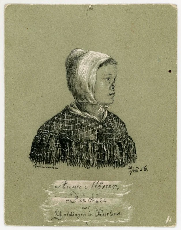
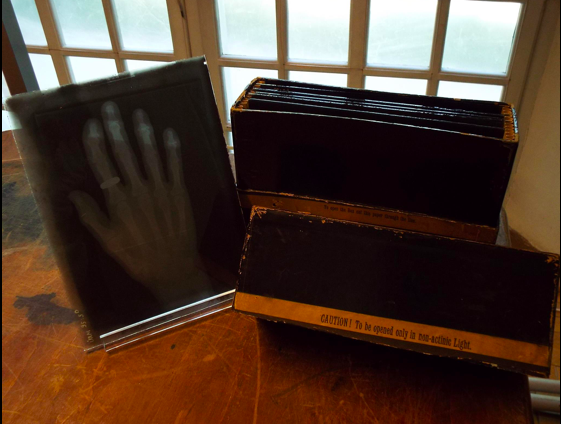
As the curator of the museum's medical collection, I was tasked with the medical sciences section of the exhibition: a skull with trepanation, rhinoplasty illustrations by Julius von Szymanowski (the father of plastic reconstructive surgery), the first X-ray negative taken at the university in the Spring of 1896, a brain tumour sample from Ludvig Puusepp's (the first professor of neurosurgery in the world) neurosurgery collection and gastroscope used in Estonian Heliobacter pylori research which contributed to the early acceptance in medicine.
Curators: Kaija-Liisa Koovit, Leili Kriis, Lea Leppik, Sirje Sisask, Kristiina Tiideberg, Tiina Vint
Julius Alfons Nikolai Szymanowski (1829-1868)
Julius Alfons Nikolai Szymanowski (1829-1868) was one of the most renowned surgeons of the 19th century. He received his medical degree from the University of Dorpat, now the University of Tartu, in 1856. While mostly forgotten in the 20th and 21st-century medical history books, he introduced modern cosmetic surgery into surgical practice. His most influential work Handbuch der operativen Chirurgie was published posthumously in 1870 and was considered the most influential surgery handbook of its time.
The museum has three original illustrations from his time as a medical student at the university.
First x-ray images
After the first publication of an x-ray and instructions on how to do it by Wilhelm Conrad Röntgen in December 1895, any respected physicists with the means to repeat the experiment did so. University of Tartu Physics Laboratory attempted theirs, with success, in early 1896. The local newspaper noted that it took half an hour and required a small gas engine with a 2.5 horsepower motor.
The museum collection has a box of the first images taken by the laboratory. While half of the original negatives are missing, the remaining six give a story of human reaction to the new invention. There are indeed photographs of bones, but four of the six negatives are of everyday items. After all, how can one confirm that the X-ray works? By taking x-rays of the content of boxes, of course!


